Grant: 001/2020
- Title: CD84/SLAMF5 cell surface signaling molecule: emerging biomarker for Celiac Disease.
- Duration: 1 Year Project
- Principal Investigator: Laura Pisapia , Istituto di Genetica e Biofisica (IGB)- CNR, Naples, Italy
- Collaboration: Antonio Rispo, Dipartimento Di Medicina Clinica e Chirurgica, Scuola di Medicina Federico II, Naples, Italy
Publications originating from the Project
- manuscript in preparation
THE STUDY
Project rationale and aims
The aim of this project was to investigate the role of the CD84/SLAMF5 protein in the immunopathogenesis of celiac disease (CD).
CD is an intolerance to gluten proteins found in wheat and other cereals such as barley and rye, manifesting in individuals carrying class II HLA genes (DQ2/DQ8 genotype) that predispose them to the condition. Although gluten and genetic predisposition are essential elements in the development of celiac disease, they are not sufficient to fully explain this pathological condition. Many mechanisms regarding the onset of the disease and the clinical assessment of the risk of illness remain unclear. To date, genetic association studies continue to identify new biomarkers potentially associated with celiac disease. Our study falls within this context.
The CD84 protein belongs to a family of proteins called SLAM/CD2, which scientific literature describes as associated with many immunological processes, such as the development, activation, and acquisition of effector functions by immune system cells involved in immunological tolerance to antigens.
We evaluated the expression of CD84 in immune cells and its functional role in the presentation of gliadin antigens in two cohorts of patients: those with active Celiac Disease (CD) and those in remission on a gluten-free diet, compared to healthy volunteers.
The study focused on two principal aims.
Aim 1 : was to assess CD84/SLAMF5 expression on total PBMCs and on specific immune cell populations in the different cohorts of subjects enrolled in the study. By using flow cytometry and multiparametric analysis, we conducted immunophenotype analysis to characterize the total surface expression of CD84 in both lymphoid and myeloid cells using a panel of monoclonal antibodies
Aim 2 : aimed to investigate the involvement of CD84 in the activation of gliadin-specific CD4+ T cells, including in cases where CD84 is depleted.
Research plan and results obtained
We enrolled in our study 74 subjects for our study classified in celiac patients (CD) with active symptoms and positive serology for anti-transglutaminase and anti-endomysium antibodies, patients in remission on a gluten-free diet (GFD) for at least 6 months with negative serology, and healthy controls (Ctrl). We purified PBMCs from blood samples derived from each subject, and used the cells for flow cytometry assays and to prepare DNA to assess the HLA-DQ genotype.
Aim 1:
We assessed the surface expression of the CD84 molecule by flow cytometry on the total PBMCs. We found that CD showed higher CD84 expression than GFD. Ctrl had lower CD84 expression than both CD and GFD patients, as showed in Figure 1, panel A and B. Multiparametric flow cytometry immunophenotyping characterized CD84 expression in lymphoid (T and B lymphocytes) and myeloid cells (monocytes and dendritic cells), revealing differences in CD84 expression among immune cells. Lymphoid cells exhibited lower and more variable CD84 expression than myeloid cells. Moreover, higher expression of CD84 was confirmed in CD compared to GFD controls groups, particularly in B cells, CD14+ monocytes, and Dendritic Cells. For some subjects we analyzed blood samples taken in the acute phase of the disease and again after at least 9 months on a gluten-free diet. We observed a significant reduction in CD84 expression on PBMCs during the transition from the acute phase to remission.
Aim 2:
To assess the role of CD84 in antigen presentation, we conducted functional assays to test its involvement in activating gliadin-specific CD4+ T cells. We used autologous B-LCLs, with different genotypes, as antigen-presenting cells (APCs) to present deamidated gliadin peptic-tryptic (PT) digest or a pool of immunodominant gliadin peptides (Pool 1-5) to CD4+ T cell lines. The tests were done with or without CD84 protein, directly blocked on the cell surface using a neutralizing CD84 antibody. CD4+ T cell activation was measured as IFN-gamma production in co-culture supernatants and by assessing CD25 on CD4 T cells, a marker of lymphocytes activation. In presence of CD84 neutralizing antibody, we observed a significant reduction in cytokine synthesis, measured by ELISA, and in CD25 surface expression on T cells, measured by flow cytometry, as shown in Figure 2 panels A and B.
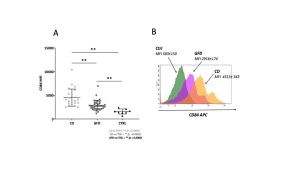
Figure 1 – Flow cytometry analysis of CD84 molecule surface expression. The surface expression of the CD84 molecule was measured on total PMBCs from celiac patients with acute CD (CD), patients on GFD(GFD) and healthy controls (CTLR). Panel A show the CD84 expression as Mean Fluorescence Intensity (MFI), for each subject analyzed. Panel B shows a representative histogram for one subject from each group with the mean of the MFI value measured and the standard error.

Figure 2. Gliadin-specific CD4+ T cells activation in the presence of anti-CD84 neutralizing antibody. The CD4+ T cells activation assay showed significant inhibition in response to gliadin digest and to a pool 1-5 of gliadin peptides, in the presence of CD84 neutralizing antibody . Panel A showed, the mean value of CD25 molecule expression reduction on CD4+ T cells, for all patients analysed . Panel B showed representative fluorescence histograms obtained by flow cytometry analysis of CD25 with and without CD84 neutralizing antibody.
Experimental design and methodologies
In our study, adult subjects in the acute phase of celiac disease (CD), patients on a gluten-free diet for at least six months, and healthy controls were enrolled and identified through clinical symptom and serology assessment.
Peripheral blood mononuclear cells (PBMCs) were isolated from patients’ blood and used for DNA and RNA extraction as well as flow cytometry analyses. DNA was extracted using the DNA Easy Kit and used for HLA-DQ genotyping. PBMCs immunophenotyping was carried out using FACSAriaIII (Becton Dickinson) instrument, and data analysis was performed by DIVA software. Anti-human-CD84 monoclonal antibody was used to detect CD84 expression on immune cells including CD4+ T cells, CD8+ T cells, B cells, monocytes, and dendritic cells.
Functional assays were conducted using B lymphoblastoid cell lines (B-LCLs) from CD patients (HLA-DQ2/DR3 genotype) as APC to present gliadin or a pool of immunodominant peptides to gliadin-specific CD4+ T cells, with or without anti-CD84 neutralizing antibody. Gliadin-specific CD4+ T cell activation was measured by Elisa test IFN-gamma , and measured CD25 expression on CD4+ T cells by flow cytometry.
Potential pitfalls and caveats
The main pitfall concerned the enrolment of healthy subjects not affected by celiac disease with HLA-DQ2 predisposing genotype, carried by 30% of the population in Campania. However, the data obtained are homogeneous and statistically reliable. A further observation concern the variability in IFN-gamma production; however, its constant reduction after CD84 neutralization clearly demonstrates CD84’s involvement in antigen presentation.
We don’t have data regarding the CD84 expression in other autoimmune disorders and no papers demonstrated its involvement in the antigen presentation, however we demonstrated the relevance of this biomarker in celiac disease. The variation from acute CD to GFD assign to CD84 a function of biomarker to monitor the compliance of therapy and the recovery from pathology.
Conclusions and discussion
The role of the CD84 protein in celiac disease has not yet been described. Our objective was to investigate its association with the immune-pathogenesis of the disease and assess its clinical usage in classifying the risk of illness and evaluating disease progression.
We assessed the expression of CD84 and demonstrated that the myeloid cells have more protein than lymphoid cells on the cell surface. We demonstrated a down-regulation of CD84 in circulating immune cells from subject in GFD and healthy controls, respect patients in acute celiac disease, and that this modulation is exclusively associated to the achievement of remission state, and excluding subject-to-subject variations.
We demonstrated the functional role of CD84 in the antigen-presentation because its depletion affects the gliadin-specific T cell activation, measured as IFN-gamma production and CD25 surface activation marker expression. We hypothesize that the amount of CD84 during antigen presentation could improves the immune response to gluten in terms of strength and duration of interaction between T cells and APCs, generating stable antigen-specific T: APC complex.
We demonstrated that CD84 can be considered a biomarker of disease.
Recognizing its predictive value for the phases and progression of celiac disease offers several advantages. Understanding its role enhances knowledge of immune activation triggered by gluten, provides a means to monitor effective therapy, and supports clinical management of patients by enabling non-invasive identification of transitions between different stages of the disease, such as from acute phase to remission. Furthermore, it presents an ideal target for developing new treatment options.